Tracking stents, instruments and embolic agents without ionizing radiation
Endovascular interventions are guided by X-ray fluoroscopy today. Magnetic particle imaging offers a radiation-free alternative with high temporal resolution, provided that instruments and implants can be made visible. In a series of studies with the radiology departments in Lübeck and Hamburg, we investigated how metallic implants interact with the MPI fields and how instruments can be marked.
Bare-metal stents generate a signal of their own in MPI. Their magnetic properties allow the position of a stent to be tracked, while the heating of large endovascular stents and stent grafts in the drive field was characterized to assess safety.
Instrument markers made of water-stable SPION coatings are simple to fabricate and remain visible in MPI even after prolonged exposure to water, which makes them suitable for preclinical catheters and stents.
Liquid embolization with MPI-based guidance allows the distribution of an embolic agent to be monitored in real time.
Bimodal OCT/MPI combines the high-resolution intravascular optical coherence tomography with the volumetric MPI information to estimate the catheter trajectory.
Left: reconstructed MPI images and centers of mass of tracked bare-metal stents (Wegner et al., International Journal of Nanomedicine 2024). Right: MPI images of a stent with water-stable instrument markers (Wegner et al., Medical Devices: Evidence and Research 2026).
References
2026
Simple-to-Fabricate and Water-Stable Instrument Markers for Preclinical Magnetic Particle Imaging and Magnetic Resonance Imaging
Abstract Purpose Intravascular optical coherence tomography (IVOCT) is a catheter based image modality allowing for high resolution imaging of vessels. It is based on a fast sequential acquisition of A-scans with an axial spatial resolution in the range of 5 to 10 μm, i.e., one order of magnitude higher than in conventional methods like intravascular ultrasound or computed tomography angiography. However, position and orientation of the catheter in patient coordinates cannot be obtained from the IVOCT measurements alone. Hence, the pose of the catheter needs to be established to correctly reconstruct the three-dimensional vessel shape. Magnetic particle imaging (MPI) is a three-dimensional tomographic, tracer-based and radiation-free image modality providing high temporal resolution with unlimited penetration depth. Volumetric MPI images are angiographic and hence suitable to complement IVOCT as a co-modality. We study simultaneous bimodal IVOCT MPI imaging with the goal of estimating the IVOCT pullback path based on the 3D MPI data. Methods We present a setup to study and evaluate simultaneous IVOCT and MPI image acquisition of differently shaped vessel phantoms. First, the inuence of the MPI tracer concentration on the optical properties required for IVOCT is analyzed. Second, using a concentration allowing for simultaneous imaging, IVOCT and MPI image data is acquired sequentially and simultaneously. Third, the luminal centerline is established from the MPI image volumes and used to estimate the catheter pullback trajectory for IVOCT image reconstruction. The image volumes are compared to the known shape of the phantoms. Results We were able to identify a suitable MPI tracer concentration of 2.5 mmol L−1 with negligible influence on the IVOCT signal. The pullback trajectory estimated from MPI agrees well with the centerline of the phantoms. Its mean absolute error ranges from 0.27mm to 0.28mm and from 0.25mm to 0.28mm for sequential and simultaneous measurements, respectively. Likewise, reconstructing the shape of the vessel phantoms works well with mean absolute errors for the diameter ranging from 0.11mm to 0.21mm and from 0.06mm to 0.14mm for sequential and simultaneous measurements, respectively. Conclusions MPI can be used in combination with IVOCT to estimate the catheter trajectory and the vessel shape with high precision and without ionizing radiation. This article is protected by copyright. All rights reserved.


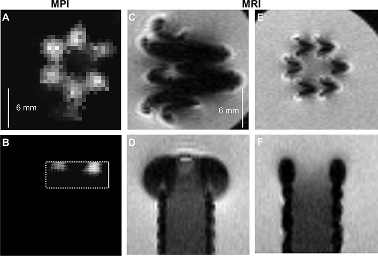 Simple-to-Fabricate and Water-Stable Instrument Markers for Preclinical Magnetic Particle Imaging and Magnetic Resonance ImagingMedical Devices: Evidence and Research, 2026
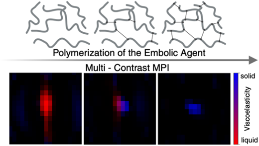 Magnetic Particle Imaging-Based Guidance of Endovascular Liquid EmbolizationACS Applied Nano Materials, 2026
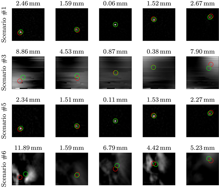 Bare-Metal Stent Tracking with Magnetic Particle ImagingInternational Journal of Nanomedicine, 2024
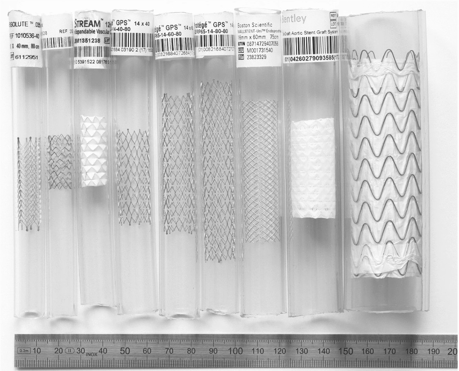 Heating of Large Endovascular Stents and Stent Grafts in Magnetic Particle Imaging—Influence of Measurement Parameters and Isocenter DistanceCardioVascular and Interventional Radiology, 2022
 Bimodal intravascular volumetric imaging combining OCT and MPIMedical Physics, 2019