MPI Head Scanner (NeuroMap)
A Stroke Surveillance System for the ICU
Time is Brain. A famous quote for stroke treatment. With 1,9 million cells dying every minute with an acute stroke, the on site detection and prevention is a very important goal. However, the treatment of stroke strongly depends on the type of stroke, namely if it is an ischemic or a hemorrhagic stroke. For this decision the patient is typically imaged by the CT within the emergency room. After treatment there is a need of further surveillance to detect a recurrance of the stroke an induced bleeding due to the treatment.
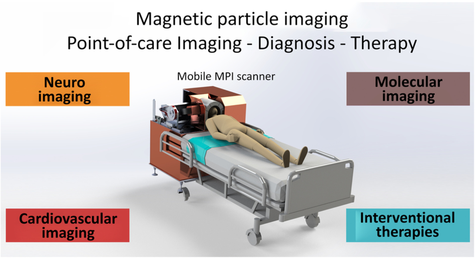
For the intensive care unit (ICU) not imaging modalities are established, as a CT has a lot of infrastructure needs and a mobile ultrasound can not image the brain within the skull. Magnetic Particle Imaging can fill this gap, as MPI systems can be build as low field systems which can work in unshielded environments and work on a standard power plug.
In 2019 I presented the first prototype of this device together with my collegues from the University Medical Center Hamburg Eppendorf IBI UKE. At the Fraunhofer IMTE we are currently redesigning the system to be mobile and match standard product specifications, and clinical requirements. As the system is so low in demands, it might even be possible to mount it within an emergency vehicle and bring the decision making direct to the patient.

To evaluate the suiteability of the system two experiments were taken out: First, a static experiment with two nanoparticle filled hemispheres containing a concentration of 965 ng/ml iron. This concentration corresponds well to the concentration expected within the brain considering both blood and brain matter. On the right hemispere a small region of 42 ml was left without nanoparticles to represent a very small stroke. This can easily be identified on the reconstructed image.
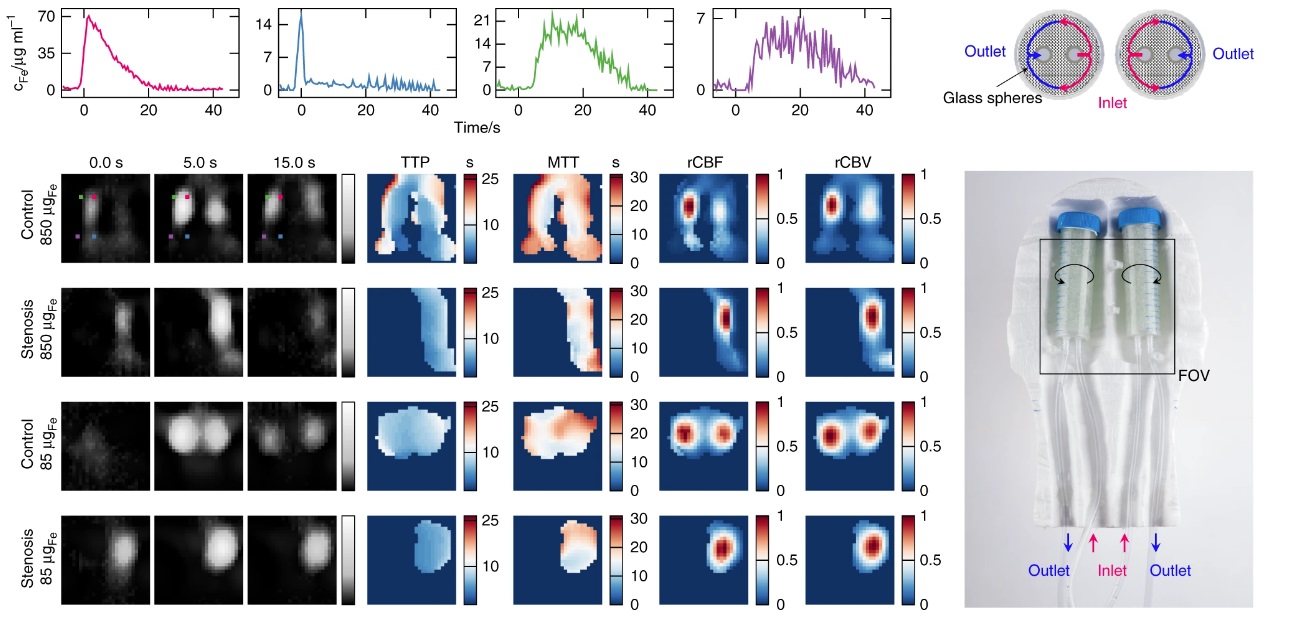
In the clinic, dynamic images are typically used to calculate perfusion parameter maps. These maps can tell the physician more about the type of stroke, the severity and the best treatment scenario. Therefor, we also did an dynamic experiment. Two perfusion chambers were constructed where an induced flow enters the chamber vom below, then passes inwards, circles around the phantom to the outer sides and exits again at the bottom. A sketch can be seen in the image below.

With the image series perfusion parameters were calculated, namely the Time to Peak map (TTP) the mean transit time (MTT), the relative cerebral blood flow (rCBF) and the relative cerebral blood volume (rCBV), which are the most relevant parameters for treatment decision.
At the Fraunhofer IMTE we are now rebuilding the proof of principle into a functional product version. For more information please contact me at matthias.graeser@imte.fraunhofer.de
References
2021
-
 Magnetic particle imaging for assessment of cerebral perfusion and ischemiaWIREs Nanomedicine and Nanobiotechnology, Sep 2021

2019
-
 Human-sized Magnetic Particle Imaging for Brain ApplicationsNature Communications, Sep 2019

2017
-
 Magnetic Particle Imaging for Real-Time Perfusion Imaging in Acute StrokeACS Nano, Sep 2017PMID: 28976180
