Receive Coil optimization for Magnetic Particle Imaging Systems
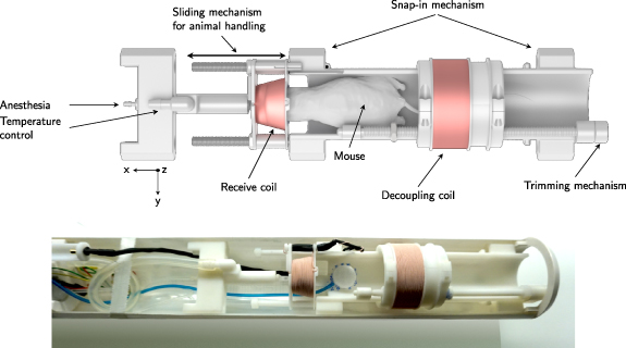
MPI systems often come with preinstalled full bore receive coils, which are good for general purpose measurements but not optimal for specific experimental applications. In this project a team of collegues, students and myself build application oriented receive coils and electronics in order to maximize SNR outcome and improve image quality. Over time the specialized coils have improved, starting from a 40mm 1D coil for mice bed, oder a 72mm 1D coil for rat beds towards full 3D coils for both rodent sizes. Due to specialized needs we also build a mouse head coil which was nested in the rat bed and a sample vial coil set for measureing system matrices or highly diluted samples.
Overview of the different application specific gradiometric coil sets designed for better SNR
Using theese coils high sensitive, high resolution and dynamic (46 volumes /s) image series were measured of a healthy mouse (left two) and a mouse suffering from a ischeamic stroke. For imaging a bolus of 10 µl perimag (micromod) was injected..
A mouse stroke study comparing two mice, one with a stroke within the left hemisphere.
From such dynamic data the perfusion parameter maps can be calculated.
Resulting perfusion parameter maps from the image series.
References
2020
Design of a head coil for high resolution mouse brain perfusion imaging using magnetic particle imaging
Magnetic particle imaging (MPI) is a novel and versatile imaging modality developing toward human application. When up-scaling to human size, the sensitivity of the systems naturally drops as the coil sensitivity depends on the bore diameter. Thus, new methods to push the sensitivity limit further have to be investigated to cope for this loss. In this paper a dedicated surface coil for mice is developed, improving the sensitivity in cerebral imaging applications. Similar to magnetic resonance imaging the developed surface coil improves the sensitivity due to the closer vicinity to the region of interest. With the developed surface coil presented in this work, it is possible to image tracer samples containing only 896 pgfe and detect even small vessels and anatomical structures within a wild type mouse model. As current sensitivity measures require a tracer system a new method for determining a sensitivity measure without this requirement is presented and verified to enable comparison between MPI receiver systems.
Monitoring Intracranial Cerebral Hemorrhage Using Multicontrast Real-Time Magnetic Particle Imaging
Magnetic particle imaging (MPI) is an innovative radiation-free tomographic imaging method providing excellent temporal resolution, contrast, sensitivity, and safety. Mobile human MPI prototypes suitable for continuous bedside monitoring of whole-brain perfusion have been developed. However, for the clinical translation of MPI, a crucial gap in knowledge still remains: while MPI can visualize the reduction in blood flow and tissue perfusion in cerebral ischemia, it is unclear whether MPI works in intracranial hemorrhage. Our objective was to investigate the capability of MPI to detect intracranial hemorrhage in a murine model. Intracranial hemorrhage was induced through the injection of collagenase into the striatum of C57BL/6 mice. After the intravenous infusion of a long-circulating MPI-tailored tracer consisting of superparamagnetic iron oxides, we detected the intracranial hemorrhage in less than 3 min and could monitor hematoma expansion in real time. Multicontrast MPI can distinguish tracers based on their physical characteristics, core size, temperature, and viscosity. By employing in vivo multicontrast MPI, we were able to differentiate areas of liquid and coagulated blood within the hematoma, which could provide valuable information in surgical decision making. Multicontrast MPI also enabled simultaneous imaging of hemorrhage and cerebral perfusion, which is essential in the care of critically ill patients with increased intracranial pressure. We conclude that MPI can be used for real-time diagnosis of intracranial hemorrhage. This work is an essential step toward achieving the clinical translation of MPI for point-of-care monitoring of different stroke subtypes.
2017
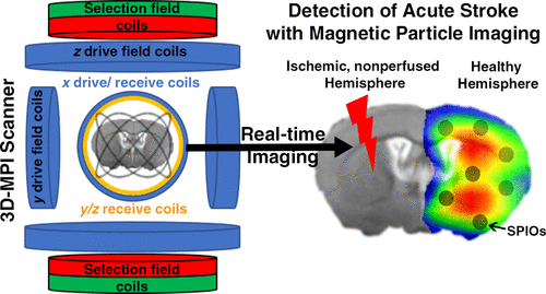
Magnetic Particle Imaging for Real-Time Perfusion Imaging in Acute Stroke


 Design of a head coil for high resolution mouse brain perfusion imaging using magnetic particle imagingPhysics in Medicine & Biology, 2020Publisher: IOP Publishing
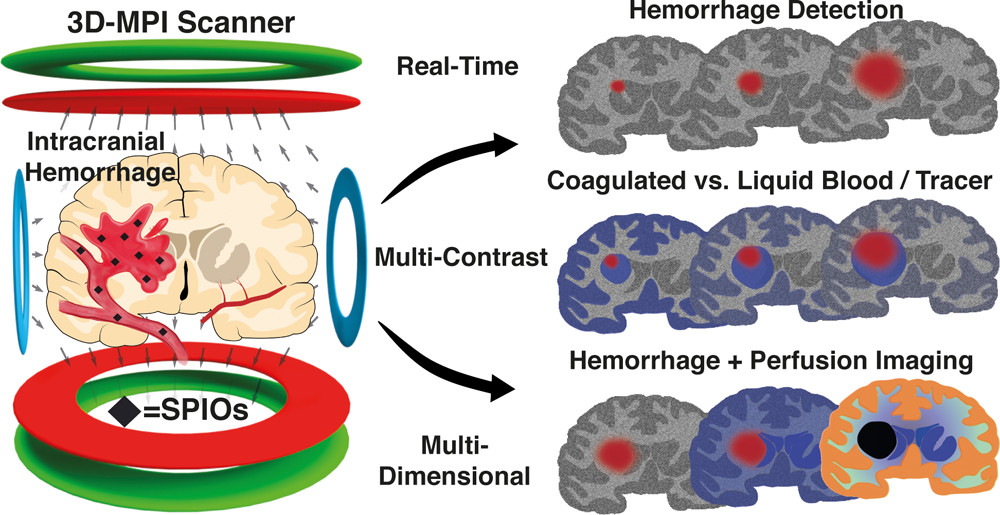 Monitoring Intracranial Cerebral Hemorrhage Using Multicontrast Real-Time Magnetic Particle ImagingACS Nano, Sep 2020

 Magnetic Particle Imaging for Real-Time Perfusion Imaging in Acute StrokeACS Nano, Sep 2017PMID: 28976180
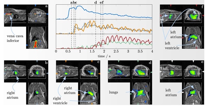 Towards picogram detection of superparamagnetic iron-oxide particles using a gradiometric receive coilScientific reports, Sep 2017
